You have symptoms that have been building for a while — heavier periods, a pressure low in your pelvis, a lower abdomen that looks different than it used to. Somewhere in the reading you have done, fibroids came up. Now you want to know whether that is actually what you have, and what it takes to find out.
The short answer is that fibroids are usually diagnosed with a pelvic ultrasound, and most women who have one get an answer the same day. The longer answer is that the ultrasound is the middle of the process rather than the beginning. What comes before it determines whether the scan gets ordered at all, and what comes after determines whether the fibroids found are actually the ones causing your symptoms.
At Seamless Medical Centers, Dr. Zagum Bhatti, Board-Certified Interventional Radiologist and Founder & Chief Medical Officer of Seamless Medical Centers, reviews fibroid imaging as a routine part of evaluation. A recurring theme among the women he sees is that they had symptoms for years before anyone imaged them, because each symptom was addressed separately and nobody connected them.
This guide walks through the diagnostic sequence: what prompts an evaluation, what a physical examination can and cannot establish, what each imaging test shows, and how a confirmed diagnosis translates into a treatment conversation.
What Usually Prompts a Fibroid Evaluation
Most fibroid diagnoses begin with a symptom rather than a screening test. There is no routine screening for fibroids, and many are found incidentally during imaging ordered for something else entirely. When they are found deliberately, it is almost always because a woman described a symptom pattern that pointed toward the uterus.
The pattern that most often prompts imaging is bleeding that has changed — heavier flow, longer periods, clots that have grown larger. Our guide to heavy menstrual bleeding covers what those thresholds are. The second common pattern is pressure rather than bleeding: a persistent fullness low in the pelvis, visible abdominal change, or new bladder and bowel symptoms, which we cover in our guide to pelvic pressure and fibroid bulk symptoms.
What matters when you describe these is specificity. “My periods are heavy” is harder to act on than “I am changing a pad every hour on my second day and passing clots bigger than a quarter.” The same applies to pressure: “my waistband stopped fitting eight months ago” carries more diagnostic weight than “I feel bloated.” Concrete detail is often what determines whether imaging gets ordered on the first visit or the third.
What a Physical Examination Can Establish
Evaluation usually begins with a history and a pelvic examination. During a bimanual examination, a clinician can feel the size, shape, and mobility of your uterus. A uterus enlarged by fibroids often feels larger than expected for your age and stage, and frequently feels irregular or lumpy rather than smooth, because fibroids distort its contour.
That finding is genuinely useful, and it is often what triggers the imaging referral. But an examination cannot tell you how many fibroids there are, exactly where they sit, or how large each one is — and those are the details that determine both whether the fibroids explain your symptoms and which treatments are appropriate. A normal examination also does not exclude fibroids: small ones, and fibroids sitting inside the uterine cavity, may not be palpable at all.
So the honest position is that a pelvic examination raises or lowers suspicion rather than settling the question. If your symptoms fit and your examination is normal, imaging is still reasonable. If you have been told your examination was normal and left it there, that is worth revisiting.
What a Pelvic Ultrasound Shows
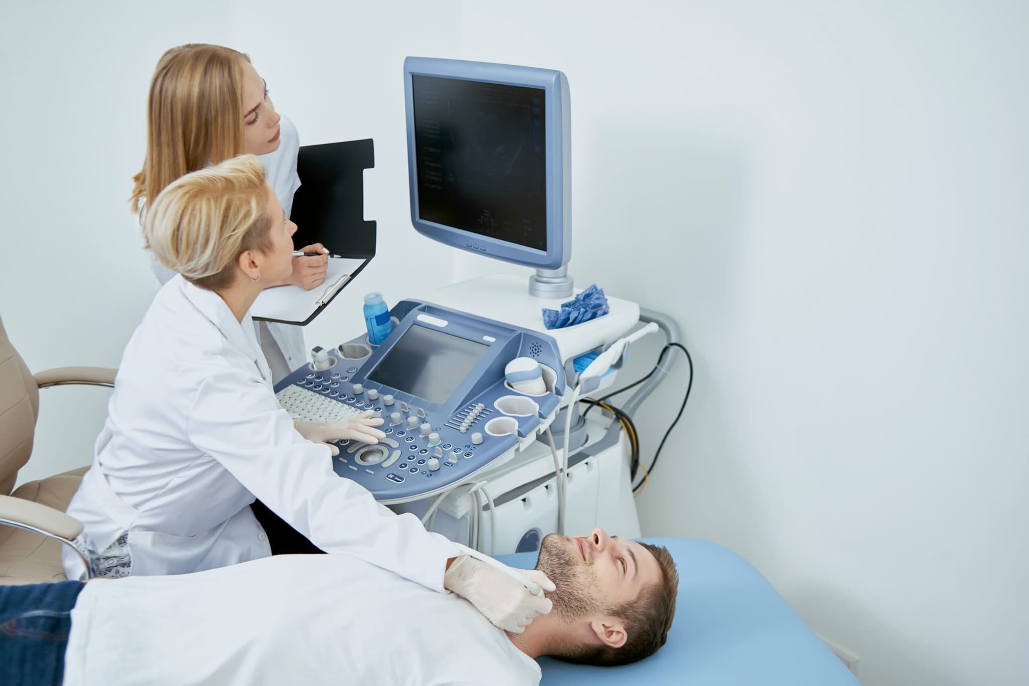
Ultrasound is the primary tool for diagnosing fibroids. It is widely available, involves no radiation, and in most cases answers the question in a single appointment. It is usually performed in two ways during the same visit — transabdominal, with the probe on your lower abdomen, and transvaginal, which places the probe closer to the uterus and gives considerably better detail of the uterine cavity and lining.
A good ultrasound report tells you four things: whether fibroids are present, how many, how large each significant one is, and where each sits relative to the uterine wall and cavity. That last detail is the one that matters most and the one most often skimmed over. Fibroids growing into the cavity behave very differently from fibroids growing outward from the outer surface, even at identical size, and the position is what explains which symptoms you have.
The report will also record the overall size of your uterus, sometimes described as bulky, which simply means larger than expected. If you want to understand what fibroids are and how they develop before your appointment, our uterine fibroids condition overview covers the background.
When MRI Is Used Instead
MRI is not the first test and most women never need one. It becomes useful in specific situations: when ultrasound cannot distinguish individual fibroids in a substantially enlarged uterus, when there are many fibroids and their relationships are unclear, when the imaging findings do not match the symptoms, or when a diagnosis of adenomyosis is being considered alongside fibroids, since the two frequently coexist and are difficult to separate on ultrasound alone.
MRI is also used specifically for treatment planning. It maps the arterial supply feeding each fibroid, which is directly relevant if uterine fibroid embolization is being considered, and it characterises fibroids in ways that help predict how they are likely to respond. If your care has reached the point of choosing between procedures, an MRI is often what makes that choice properly informed rather than approximate.
It is worth knowing this in advance, because being told you need an MRI after an ultrasound can feel like escalation. In this context it usually is not. It generally means the plan is moving from establishing what you have toward deciding what to do about it.
One practical note about scheduling. Ultrasound findings can vary slightly depending on where you are in your cycle, because the uterine lining thickens and thins across the month. If your imaging is specifically assessing the lining rather than the fibroids themselves, your clinician may ask you to attend at a particular point in your cycle. For straightforward fibroid assessment this rarely matters, and you should not delay a scan waiting for a perfect window. If nobody mentions timing, it is reasonable to ask whether it applies in your case rather than assuming it does.
The Tests That Run Alongside Imaging
Imaging identifies fibroids. It does not tell you what the bleeding has done to you, and it does not exclude the other conditions that produce similar symptoms. That is why a thorough evaluation pairs imaging with blood work and, in some cases, further assessment of the uterine lining.
- A complete blood count, which establishes whether the bleeding has caused anemia.
- Iron studies including ferritin, which detect depleted iron stores before hemoglobin falls — a normal blood count alone does not rule out iron deficiency.
- Thyroid function testing, since thyroid disorders are a correctable cause of heavy bleeding that is simple to exclude.
- Assessment of clotting function where personal or family history suggests a bleeding disorder.
- Evaluation of the uterine lining where the bleeding pattern warrants it, particularly with new symptoms after age forty.

Are You a Candidate for PAD Treatment?
If you answer "yes" to any of these questions, you may be a candidate for this treatment.
Call Now — (409) 213-9575Question 1 of 4
Have you been diagnosed with Peripheral Artery Disease (PAD) by a doctor?
Prefer to talk? Call (409) 213-9575
Your information is protected under HIPAA. We will never share your data.
The iron result is the one most often overlooked. Fibroids can be confirmed and treatment planned while the fatigue that brought a woman in gets no attention at all, because her hemoglobin came back within range. Our guide to heavy bleeding and anemia explains why ferritin is the more sensitive measure and why it is worth asking for by name.
When Fibroids Are Found but May Not Be the Cause
Fibroids are common, and finding them does not automatically mean they explain your symptoms. This is the step most easily skipped, and skipping it leads to treating the wrong thing. A small fibroid on the outer surface of the uterus is unlikely to be responsible for very heavy bleeding. A woman with pelvic pain that worsens through the day on standing may have fibroids and also have a separate condition producing the pain.
The question a good evaluation answers is not only “are there fibroids” but “do these fibroids, in these positions, at these sizes, account for these symptoms.” Where the answer is no, or only partly, the workup should continue rather than stop. Adenomyosis, polyps, hormonal causes, and clotting disorders all produce overlapping pictures and are managed differently.
It is worth naming the concern that sits behind a lot of searching about diagnosis. This is not a reason to assume the worst — the overwhelming majority of fibroids are benign and remain so — but it is a reason to have persistent symptoms properly evaluated rather than assuming an incidental finding explains everything you are experiencing.
From Diagnosis to a Treatment Conversation
Once fibroids are confirmed and matched to your symptoms, the conversation shifts to what you want to do. Not every fibroid needs treating; fibroids found incidentally in someone without symptoms are usually monitored rather than treated. Where symptoms do warrant treatment, the options range from medical management through to procedures that shrink or remove the fibroids, including uterine fibroid embolization, myomectomy, and hysterectomy.
Which fits depends on your symptoms, the size and position of the fibroids, your age, and your fertility plans. Our overview of how uterine fibroid embolization works and who it helps explains the minimally invasive option in detail, and our comparisons of UFE and hysterectomy and UFE and myomectomy set the trade-offs side by side.
Bring your imaging to any consultation about treatment. A specialist reviewing the actual scan can tell you far more about your options than one working from a summary, and it frequently saves a repeat appointment.
Frequently Asked Questions About Fibroid Diagnosis
How do I know if I have fibroids?
You cannot know from symptoms alone, because heavy bleeding and pelvic pressure have several possible causes. A pelvic ultrasound is the usual way fibroids are confirmed, and it is reasonable to ask for one if your periods have become heavier or longer, or if you have persistent pelvic pressure.
Can a pelvic exam detect fibroids?
A pelvic examination can suggest fibroids by identifying a uterus that feels enlarged or irregular, and that finding often prompts imaging. It cannot establish how many fibroids there are, their size, or their position, and a normal examination does not rule fibroids out.
What does a fibroid feel like?
Fibroids themselves are not usually felt directly. What you notice is their effect — heavier bleeding, a persistent fullness or heaviness low in the pelvis, or in some cases a firm mass in the lower abdomen. A clinician performing a bimanual examination may feel an enlarged, irregularly shaped uterus.
Do I need an MRI for fibroids?
Most women do not. MRI is used when ultrasound cannot give enough detail, when many fibroids make individual assessment difficult, when adenomyosis is also being considered, or for treatment planning — particularly to map the blood supply before embolization.
What are the early signs of fibroids?
Fibroids often produce no symptoms early on and are found incidentally. When symptoms do appear, a change from your own normal is the most useful signal: periods that have become heavier or longer, clots that have grown larger, or a new sense of pelvic pressure.
Can fibroids be diagnosed without imaging?
Not reliably. History and examination can raise suspicion, but imaging is what confirms fibroids and establishes their number, size, and position — the details that determine whether they explain your symptoms and which treatments are appropriate.
What should I bring to a fibroid consultation?
Bring any prior imaging, ideally the actual images rather than only the report, along with recent blood work. A short record of your bleeding pattern — how often you change protection on your heaviest day, clot size, and how many days you bleed — makes the consultation considerably more productive.
Is UFE appropriate if I want to become pregnant?
Uterine fibroid embolization is generally not recommended as a first-line option for women planning future pregnancy; myomectomy is typically preferred in that situation. Raise your fertility plans at consultation so the recommendation reflects them.
Talk to a Specialist About Your Symptoms
If you have symptoms that point toward fibroids and want a clear diagnosis, Contact Seamless Medical Centers to discuss your options. An evaluation establishes what is driving your symptoms and what can reasonably be done about it.
Phone: 409-213-9575
Address: 3300 Jimmy Johnson Blvd, Suite #130, Port Arthur, Texas 77642
Medical Disclaimer
Individual results may vary. This information is for educational purposes only and should not replace professional medical advice. Treatment decisions should be made in consultation with qualified healthcare providers.
Published by Seamless Medical Centers | Clinical information reflects the expertise of Dr. Zagum Bhatti, MD, Board-Certified Interventional Radiologist, Founder of Seamless Medical Centers.
Why Choose Seamless Medical Centers?
- Minimally Invasive: Most procedures require only a small incision and are performed as outpatient services.
- Expert Care: Board-certified interventional radiologists with extensive training and experience.
- Faster Recovery: Less downtime compared to traditional surgery, getting you back to your life sooner.
- Advanced Technology: State-of-the-art imaging and treatment equipment for precise, effective care.
- Patient-Centered: Personalized treatment plans tailored to your unique needs and goals.
Published by Seamless Medical Centers
Clinical information on this site is based on the expertise of Zagum Bhatti, M.D. — Board Certified in Interventional Radiology/Diagnostic Radiology (American Board of Radiology), dual-fellowship-trained (Neuroradiology; Vascular & Interventional Radiology, Rush University Medical Center), former Assistant Professor of VIR at UTHealth Houston, and Founder & Chief Medical Officer of Seamless Medical Centers.
Why Choose Seamless Medical Centers?
- Minimally Invasive: Most procedures require only a small incision and are performed as outpatient services.
- Expert Care: Board-certified interventional radiologists with extensive training and experience.
- Faster Recovery: Less downtime compared to traditional surgery, getting you back to your life sooner.
- Advanced Technology: State-of-the-art imaging and treatment equipment for precise, effective care.
- Patient-Centered: Personalized treatment plans tailored to your unique needs and goals.