You have the report in front of you. It says your uterus is bulky. It lists measurements in centimetres, describes something as submucosal, mentions the anterior wall, and ends with a recommendation you do not fully follow. Nobody walked you through it, and your follow-up appointment is three weeks away.
Imaging reports are written for the clinician who ordered them, not for you. That is not an oversight — it is what they are for. But the information in yours is the single most useful thing you have when deciding what to do about your fibroids, because position and size determine which symptoms you have and which treatments are realistic. Being able to read it changes the conversation you can have.
At Seamless Medical Centers, Dr. Zagum Bhatti, Board-Certified Interventional Radiologist and Founder & Chief Medical Officer of Seamless Medical Centers, reviews fibroid imaging as the starting point of every evaluation. Reading scans is the core of interventional radiology, and it is often where a treatment conversation genuinely begins rather than where it ends.
This guide covers what an ultrasound shows and how it is performed, what each part of the report means, when MRI adds something ultrasound cannot, and — just as importantly — what imaging cannot tell you.
How a Fibroid Ultrasound Is Performed
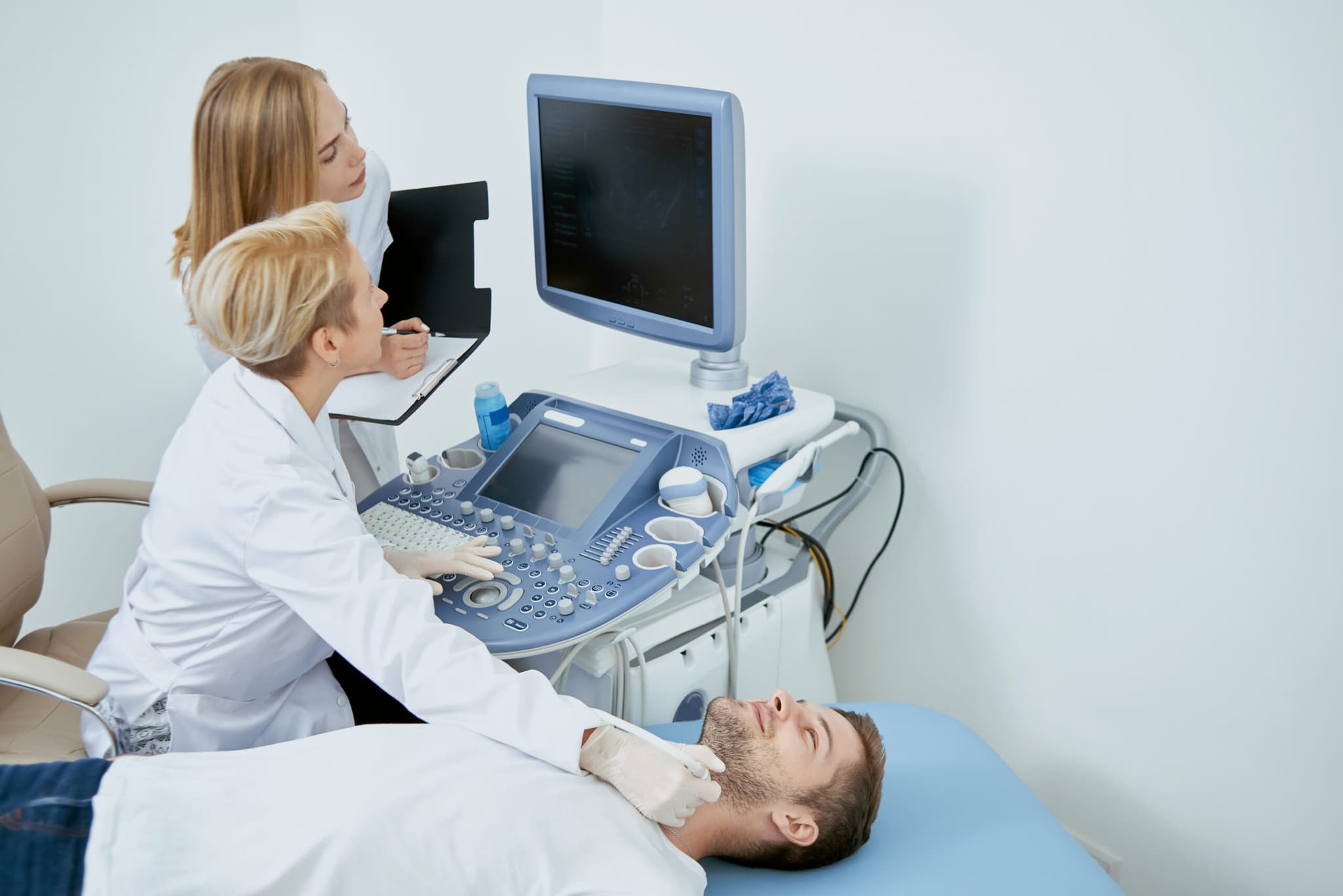
A pelvic ultrasound for fibroids is usually done in two parts during the same appointment. The transabdominal portion places the probe on your lower abdomen and gives a wide view, which is useful for seeing the overall size and shape of the uterus and for picking up larger fibroids sitting toward the outside. It generally requires a reasonably full bladder, which lifts the uterus into better view.
The transvaginal portion places a slim probe inside the vagina, much closer to the uterus. Because it sits closer, it produces considerably better resolution of the uterine cavity, the lining, and smaller fibroids — particularly those growing into the cavity, which are frequently the ones causing heavy bleeding and are the easiest to miss from the abdomen. It is usually done with an empty bladder, which is why you may be asked to empty it partway through the appointment.
Both views matter, and a scan using only one gives an incomplete picture. If your report describes only a transabdominal study and your main symptom is heavy bleeding, that is a reasonable thing to raise, because the cavity is exactly where the relevant detail sits and it is exactly what the abdominal view sees least well.
Reading the Measurements on Your Report
Your report will give the dimensions of your uterus, usually as three measurements in centimetres, and it may describe the uterus as bulky or enlarged. Bulky is a descriptive term rather than a diagnosis — it simply means the uterus measures larger than expected. Fibroids and adenomyosis are the two most common explanations, and the report may or may not distinguish between them.
Individual fibroids are then listed with their own measurements and locations. A well-written report numbers them and describes each separately. A less detailed one may summarise, noting multiple fibroids with only the largest measured. If yours is the second kind and you are considering treatment, it is reasonable to ask for a more detailed read, because a summary does not carry enough information to plan a procedure.
Your report may also comment on the endometrium, meaning the lining of the uterus, usually with a thickness measurement. This is recorded because the lining is where bleeding originates, and because a lining that appears unusually thick may prompt further assessment independent of any fibroids present. If your report mentions the endometrium and nobody has explained why, that is worth asking about directly rather than assuming it relates to the fibroids.
Resist the urge to compare your numbers against thresholds you find online. Size in isolation predicts very little. A modest fibroid pressing directly on the bladder can be considerably more disruptive than a larger one growing outward with room to expand. What the measurements are genuinely useful for is tracking change over time, which is why keeping copies of every scan matters more than interpreting any single one.
Comparison between scans comes with its own caveat. Measurements vary somewhat depending on who performed the scan, which plane the fibroid was measured in, and where you were in your cycle. A difference of a few millimetres between two reports is often measurement variation rather than genuine growth. Meaningful change is change that is clear, consistent, and visible across more than one interval — which is another argument for keeping your images rather than relying on remembered numbers.
What the Position of a Fibroid Means on Your Report
Position is the part of the report that explains your symptoms, and it is the part most worth understanding. Fibroids are described by where they sit in relation to the wall of the uterus, and each position produces a recognisably different pattern.
- Submucosal. Growing into the uterine cavity, beneath the lining. These are disproportionately associated with heavy and prolonged bleeding, because they increase the surface area that sheds each cycle and interfere directly with the cavity. They can be small and still cause significant symptoms.
- Intramural. Sitting within the muscular wall itself. The most common position. Depending on size, they can both increase bleeding and contribute to pressure, and they interfere with the uterus contracting down uniformly at the end of a period.
- Subserosal. Growing outward from the outer surface. These tend to produce pressure symptoms rather than bleeding, because they press on neighbouring structures while leaving the cavity largely undisturbed.
- Pedunculated. Attached to the uterus by a stalk, either inside the cavity or on the outer surface. Position and mobility both matter here, and this is one of the situations where the treatment recommendation may differ from the usual pattern.
- Anterior, posterior, and fundal. These describe which part of the uterus a fibroid sits on — the front, the back, or the top. Anterior fibroids sit against the bladder and tend to produce urinary symptoms. Posterior fibroids sit toward the bowel and lower back. Fundal fibroids sit at the top, often with more room before they press on anything.
Read alongside your symptoms, this is often the moment the report starts making sense. A submucosal fibroid on a report belonging to someone with very heavy periods is a coherent picture. An anterior fibroid on a report belonging to someone who has been investigated repeatedly for urinary problems is another — and our guide to fibroid-related urinary symptoms covers why that pattern is so often mistaken for recurrent infection.
If your symptoms are pressure and abdominal change rather than bleeding, subserosal and larger intramural fibroids are usually the relevant ones, and our guide to pelvic pressure and fibroid bulk symptoms explains how position translates into what you actually feel.
When MRI Adds Something Ultrasound Cannot
Most women never need an MRI, and being offered one does not mean something concerning has been found. MRI becomes useful in a defined set of situations, and all of them are about detail rather than severity.
The first is a substantially enlarged uterus with many fibroids, where ultrasound struggles to distinguish individual fibroids from one another and cannot reliably establish how many there are or exactly where each sits. The second is when the imaging and the symptoms do not match — when a scan shows little that would explain significant bleeding, for instance. The third is when adenomyosis is being considered alongside fibroids, which is common, since the two frequently coexist and are genuinely difficult to separate on ultrasound.

Are You a Candidate for PAD Treatment?
If you answer "yes" to any of these questions, you may be a candidate for this treatment.
Call Now — (409) 213-9575Question 1 of 4
Have you been diagnosed with Peripheral Artery Disease (PAD) by a doctor?
Prefer to talk? Call (409) 213-9575
Your information is protected under HIPAA. We will never share your data.
It is also worth knowing what the appointment involves, since MRI is less familiar than ultrasound. It takes longer, generally in the region of half an hour to an hour, and requires lying still inside a scanner that is enclosed and loud. Contrast material is sometimes given through a vein to show blood flow more clearly, which is particularly relevant when the arterial supply to fibroids is being assessed. If enclosed spaces are difficult for you, say so when the scan is booked rather than on the day, because there are usually options.
The fourth reason is treatment planning, and it is the one most relevant if you are weighing a procedure. MRI maps the arterial supply feeding each fibroid, which is directly relevant to embolization, and it characterises fibroid tissue in ways that help predict how a fibroid is likely to respond to treatment. An MRI at this stage usually signals that the plan is moving from establishing what you have toward deciding what to do about it.
What Imaging Cannot Tell You
Imaging identifies fibroids and describes them precisely. It does not tell you what your bleeding has done to your iron stores, which is why blood work runs alongside it and why ferritin is worth asking for by name. It does not exclude the hormonal and clotting causes that produce overlapping symptoms. And it does not, on its own, establish that the fibroids it found are the reason you feel unwell.
That last point deserves emphasis, because fibroids are common enough that finding them is not the same as explaining your symptoms. A small subserosal fibroid is unlikely to be behind very heavy bleeding, and a woman may have fibroids on her scan and a separate condition producing her pain. Our guide to how fibroids are diagnosed covers the full evaluation sequence and the step where imaging findings are matched against symptoms.
It is worth naming the anxiety that a scan report can produce. This is not a reason to assume the worst — the overwhelming majority of fibroids are benign and remain so — but unfamiliar terminology on a page you were not given help interpreting is a reasonable thing to bring back to the clinician who ordered it and ask about directly.
How Your Imaging Shapes Your Options
Once position and size are established, the treatment conversation becomes considerably more specific. Submucosal fibroids inside the cavity can sometimes be removed through the cervix without an external incision. Uterine fibroid embolization works by blocking the arterial supply so fibroids shrink, and because the mechanism is volume reduction it addresses both bleeding and pressure, with the uterus preserved. Myomectomy removes fibroids surgically. Hysterectomy removes the uterus.
Which of these is appropriate depends on what your imaging shows, what your symptoms are, and what you want for your fertility. Our overview of how uterine fibroid embolization works and who it helps explains candidacy in detail, and our comparisons of UFE and hysterectomy and UFE and myomectomy set the differences out side by side.
Bring the actual images to any consultation, not only the written report. A specialist reviewing the images directly can tell you considerably more about your options than one working from a summary, and it frequently avoids a repeat scan.
Frequently Asked Questions About Fibroid Imaging
What does a bulky uterus mean on an ultrasound report?
Bulky means the uterus measures larger than expected. It is a description rather than a diagnosis, and fibroids and adenomyosis are the most common explanations. It should be read alongside your symptoms by the clinician who ordered the scan.
Do I need an MRI for fibroids?
Most women do not. MRI is used when ultrasound cannot distinguish individual fibroids in an enlarged uterus, when the imaging does not match the symptoms, when adenomyosis is also being considered, or for treatment planning — particularly to map the blood supply before embolization.
What is the difference between submucosal, intramural, and subserosal fibroids?
The terms describe position. Submucosal fibroids grow into the uterine cavity and are strongly associated with heavy bleeding. Intramural fibroids sit within the muscular wall. Subserosal fibroids grow outward from the outer surface and tend to produce pressure rather than bleeding.
Is a transvaginal ultrasound necessary?
For assessing fibroids it usually adds meaningful detail, particularly of the uterine cavity and lining where smaller fibroids affecting bleeding are easiest to miss from the abdomen. If your main symptom is heavy bleeding and only an abdominal scan was performed, it is reasonable to ask about this.
Can an ultrasound tell how serious a fibroid is?
It establishes size and position, which together explain a great deal about your symptoms. Size alone predicts relatively little, since a smaller fibroid in a sensitive position can be more disruptive than a larger one with room to grow outward.
Should I get copies of my imaging?
Yes. Keep the images themselves rather than only the report. They allow comparison over time, they let a specialist assess your options directly, and they often prevent a repeat scan when you seek a second opinion.
Is UFE appropriate if I want to become pregnant?
Uterine fibroid embolization is generally not recommended as a first-line option for women planning future pregnancy; myomectomy is typically preferred in that situation. Raise your fertility plans at consultation so the recommendation reflects them.
Talk to a Specialist About Your Symptoms
If you have imaging and want to understand what it means for your treatment options, Contact Seamless Medical Centers to discuss your options. An evaluation establishes what is driving your symptoms and what can reasonably be done about it.
Phone: 409-213-9575
Address: 3300 Jimmy Johnson Blvd, Suite #130, Port Arthur, Texas 77642
Medical Disclaimer
Individual results may vary. This information is for educational purposes only and should not replace professional medical advice. Treatment decisions should be made in consultation with qualified healthcare providers.
Published by Seamless Medical Centers | Clinical information reflects the expertise of Dr. Zagum Bhatti, MD, Board-Certified Interventional Radiologist, Founder of Seamless Medical Centers.
Why Choose Seamless Medical Centers?
- Minimally Invasive: Most procedures require only a small incision and are performed as outpatient services.
- Expert Care: Board-certified interventional radiologists with extensive training and experience.
- Faster Recovery: Less downtime compared to traditional surgery, getting you back to your life sooner.
- Advanced Technology: State-of-the-art imaging and treatment equipment for precise, effective care.
- Patient-Centered: Personalized treatment plans tailored to your unique needs and goals.
Published by Seamless Medical Centers
Clinical information on this site is based on the expertise of Zagum Bhatti, M.D. — Board Certified in Interventional Radiology/Diagnostic Radiology (American Board of Radiology), dual-fellowship-trained (Neuroradiology; Vascular & Interventional Radiology, Rush University Medical Center), former Assistant Professor of VIR at UTHealth Houston, and Founder & Chief Medical Officer of Seamless Medical Centers.
Why Choose Seamless Medical Centers?
- Minimally Invasive: Most procedures require only a small incision and are performed as outpatient services.
- Expert Care: Board-certified interventional radiologists with extensive training and experience.
- Faster Recovery: Less downtime compared to traditional surgery, getting you back to your life sooner.
- Advanced Technology: State-of-the-art imaging and treatment equipment for precise, effective care.
- Patient-Centered: Personalized treatment plans tailored to your unique needs and goals.