
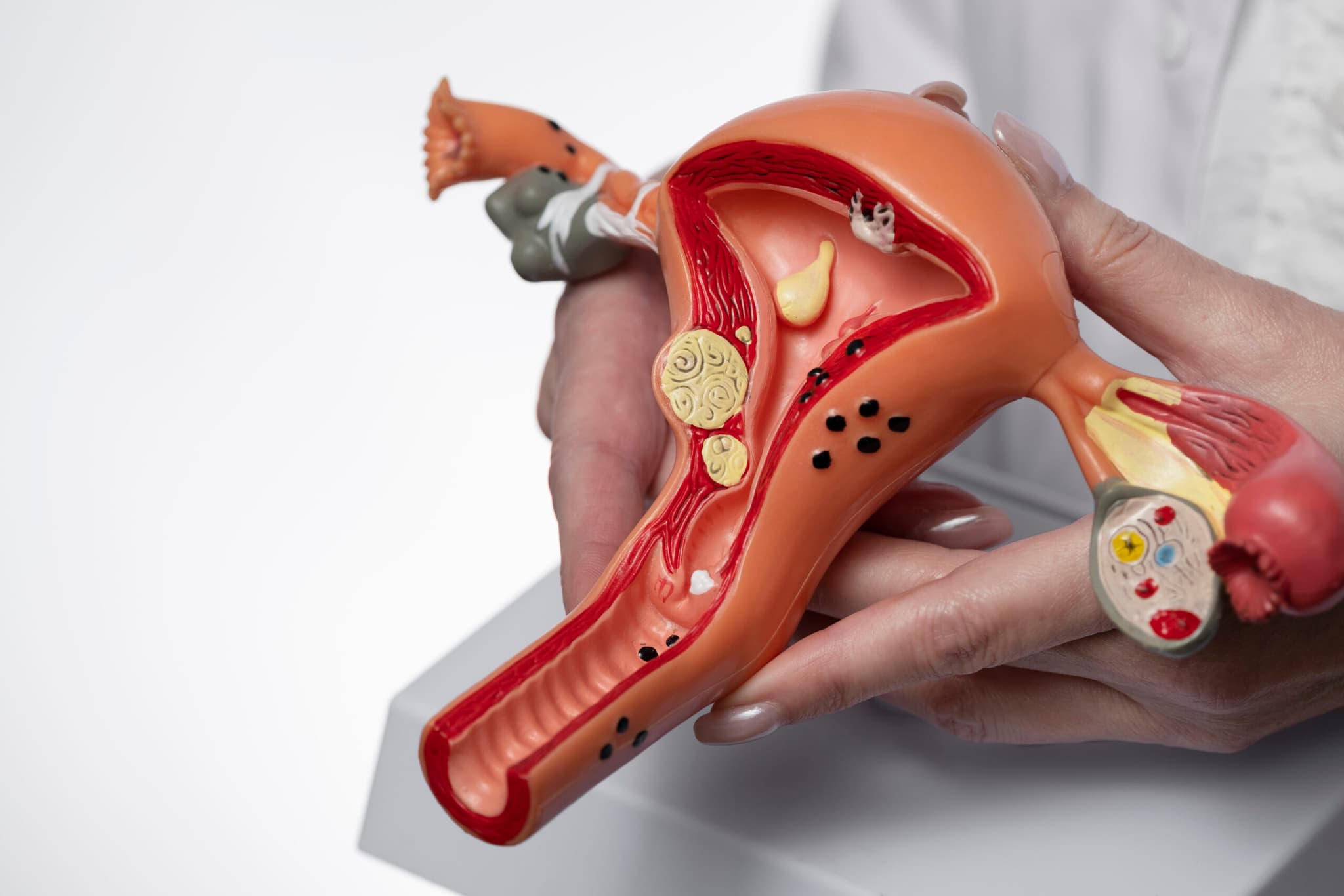
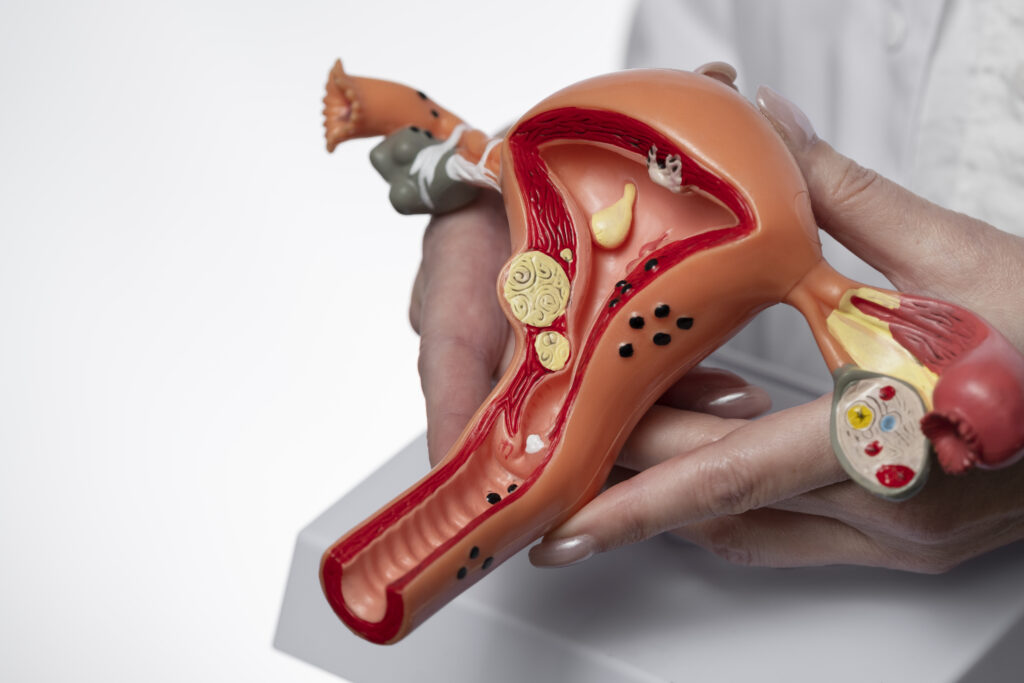
You have been told you have fibroids, and somewhere between the appointment and now the question arrived anyway. Possibly a scan report used a word you did not recognise. Possibly you have been managing symptoms for years and started wondering what else might explain them. Either way you want a straight answer rather than a reassuring one.
The straight answer is this. Fibroids are benign growths of uterine muscle tissue, and they do not turn into cancer. There is a separate, rare cancer of the uterine muscle called leiomyosarcoma, and because it arises in the same tissue it can resemble a fibroid on imaging. It is not a fibroid that became malignant — it is a different condition from the outset. That distinction is the whole substance of this question, and it is the part most sources skip.
At Seamless Medical Centers, Dr. Zagum Bhatti, Board-Certified Interventional Radiologist and Founder & Chief Medical Officer of Seamless Medical Centers, this question comes up in most consultations even when nobody raises it out loud. It deserves a direct answer, which is what follows — including the parts that are less tidy than a simple reassurance.
Fibroids Are Benign, and They Do Not Become Malignant
A fibroid is an overgrowth of the smooth muscle tissue that makes up the wall of the uterus, wrapped in fibrous material and fed by its own blood supply. They are extremely common — most women develop at least one during their reproductive years — and the overwhelming majority cause either no symptoms at all or symptoms that are a quality-of-life problem rather than a health threat.
It helps to know why the word benign is used so confidently here. A fibroid is defined by what its cells actually are when examined — orderly smooth muscle tissue, growing in a contained mass, without the features that characterise malignant tissue. This is not a probabilistic reassurance about fibroids as a category; it is what the diagnosis means. A growth that turned out to have different cellular features would not be called a fibroid.
The transformation model many women have in mind, where a benign growth gradually turns into a malignant one over time, does not describe what happens here. Having fibroids does not raise your risk of uterine cancer, and having had them for many years does not accumulate risk. A fibroid you have carried for a decade is not more dangerous for having been there a decade.
This also means that a fibroid growing is not, in itself, a warning sign. Growth is what fibroids do, it is slow and uneven and varies enormously between women, and our guide to how fast fibroids grow covers what is and is not known about it. Size, similarly, is a poor predictor of almost everything — our guide to fibroid size and what it actually determines explains why position matters more.
The Separate Condition, and Why It Causes Confusion
Leiomyosarcoma is a cancer that arises in the smooth muscle of the uterus. It is rare — substantially rarer than fibroids, which are among the most common conditions in women — and it is a distinct disease rather than a stage of one. The confusion exists because both develop in the same tissue and can therefore look similar on a scan.
It is most often found in women past menopause, though not exclusively, and it tends to behave differently from a fibroid: growing quickly rather than slowly, and frequently appearing at a point in life when fibroids would ordinarily be becoming less active rather than more. Those behavioural differences are what raise or lower suspicion clinically, far more than any single feature on a scan.
It is worth holding the proportions in mind while reading this. Fibroids are ordinary; this condition is rare. Most women reading this page have fibroids and will simply have fibroids. The reason it merits a clear explanation rather than a passing mention is that a vague answer leaves the question open, and an open question is considerably more corrosive than an honest one.
Why Imaging Cannot Fully Settle It
Here is the part usually left out, and it is the reason this page exists. Ultrasound and MRI can characterise a growth in real detail — size, position, blood supply, tissue characteristics — and MRI in particular can identify features that make one explanation more or less likely. What no scan can do is provide a definitive answer, because that requires examining the tissue itself under a microscope. Our guide to what your fibroid imaging shows covers what a report can and cannot establish.
This is not a gap in your particular care or a limitation of a particular clinic. It is inherent to the two conditions arising in the same tissue. In practice it rarely changes anything: the overwhelming likelihood in any given case is a fibroid, and treatment proceeds on that basis. It matters in the small number of situations where the clinical picture is genuinely unusual, and knowing about it in advance means that if your clinician suggests a different approach, you understand why rather than being alarmed by it.
It is also worth being clear about what happens in the ordinary case, because the paragraph above can read as more ominous than the reality warrants. For the vast majority of women, imaging shows fibroids, the picture is entirely consistent, nobody raises any other possibility, and treatment proceeds on that basis without the question ever arising. The uncertainty described here is real in principle and almost never operative in practice, which is precisely why it belongs in a paragraph rather than a warning.
The practical upshot is that clinical judgement carries the weight here — how the growth is behaving, at what age, alongside which symptoms. That is a more reliable guide than any single scan feature, and it is why a specialist reviewing your actual imaging and history is worth more than any amount of reading.
What Would Prompt Closer Attention
A short list of situations leads clinicians to look harder rather than proceed on assumption. Rapid growth over a short period, particularly where growth is clearly out of keeping with the slow pattern fibroids usually follow. Growth after menopause, when declining hormones would ordinarily make fibroids less active. New or unusual symptoms that do not fit the fibroid picture, including bleeding after menopause has completed — which is always a reason for prompt assessment, whatever the suspected cause. Our guide to how fibroids are diagnosed sets out the evaluation.
Worth adding to that list is the situation where symptoms and imaging simply do not match — significant symptoms with findings that do not account for them, or the reverse. That mismatch is common and almost always has an ordinary explanation, frequently adenomyosis or a second condition alongside the fibroids. It earns further evaluation rather than a procedure, which is the same conclusion the rest of this cluster keeps arriving at from different directions.

Are You a Candidate for PAD Treatment?
If you answer "yes" to any of these questions, you may be a candidate for this treatment.
Call Now — (409) 213-9575Question 1 of 4
Have you been diagnosed with Peripheral Artery Disease (PAD) by a doctor?
Prefer to talk? Call (409) 213-9575
Your information is protected under HIPAA. We will never share your data.
If any of those apply to you, the appropriate response is an appointment, not alarm. Each of them has ordinary explanations that are far more likely than the rare one, and the point of assessment is to establish which applies rather than to confirm a fear. What none of them warrants is waiting to see whether things settle.
And if none of them applies — if you have fibroids that are behaving the way fibroids behave, producing the symptoms fibroids produce — then the honest answer to the question that brought you here is that this is not what you are dealing with, and the more useful question is what to do about the symptoms you actually have.
What This Means for Treatment
For the great majority of women, this question does not change the treatment conversation at all. Where fibroids are confirmed and symptoms warrant treatment, the options are the usual ones: medical management, uterine fibroid embolization, myomectomy, and hysterectomy. Our overview of how UFE works and who it helps covers candidacy, and our comparisons of UFE and hysterectomy and UFE and myomectomy set out the alternatives.
There is one genuine exception, and it is worth stating plainly. Where malignancy is actively suspected, embolization is not the appropriate path. Procedures that shrink or remove growths without producing tissue for examination are not how you answer a diagnostic question — that requires a diagnosis first, and a treatment plan built on it. Any specialist should tell you this unprompted; it belongs in the same category as the other honest limitations set out in our guide to UFE side effects and who it suits less well.
If the question has been weighing on you, it is worth raising it out loud at your next appointment rather than carrying it. Clinicians are entirely used to it, it takes very little time to address in the context of your own imaging and history, and a specific answer about your situation settles it far more effectively than any general reading can. Women frequently leave consultations having discussed everything except the thing that worried them most.
In practice this means that a thorough evaluation before treatment serves two purposes at once. It establishes whether your fibroids explain your symptoms, which is what determines whether treating them will help. And it establishes that the clinical picture is what it appears to be. Both are reasons to want the assessment done properly rather than quickly, and both are questions you are entitled to ask directly.
Frequently Asked Questions
Can fibroids turn into cancer?
No. Fibroids are benign growths of uterine muscle and do not transform into malignant ones. Leiomyosarcoma is a separate, rare cancer that arises in the same tissue and can resemble a fibroid on imaging, but it is a distinct condition rather than a fibroid that changed.
Do fibroids increase my risk of uterine cancer?
Having fibroids does not raise your risk of uterine cancer. Nor does having had them for many years accumulate risk over time.
Does a fibroid growing mean something is wrong?
Growth is normal fibroid behaviour and is usually slow and uneven. What prompts closer attention is growth that is rapid and clearly out of keeping with that pattern, or growth occurring after menopause when fibroids would ordinarily become less active.
Can an ultrasound tell whether a growth is cancerous?
Imaging can characterise a growth in detail and identify features that make one explanation more or less likely, and MRI adds considerably to that. No scan provides a definitive answer, because that requires examining the tissue itself. In practice the overwhelming likelihood is a fibroid, and clinical judgement guides the rest.
Should I have my fibroids removed just to be safe?
Fibroids causing no symptoms are usually monitored rather than treated, and being benign is precisely why that approach is reasonable. Treatment decisions are guided by symptoms rather than by precaution against a condition fibroids do not become.
What symptoms should prompt prompt evaluation?
Bleeding after menopause has completed, rapid change in size, and new symptoms that do not fit your usual pattern. Each has ordinary explanations that are far more likely, and each is a reason to be seen rather than to wait.
Is UFE appropriate if malignancy has been raised as a possibility?
No. Where malignancy is actively suspected, the priority is establishing a tissue diagnosis rather than performing a procedure that shrinks a growth without producing tissue for examination. Treatment planning follows diagnosis, not the other way around.
Is UFE appropriate if I want to become pregnant?
Uterine fibroid embolization is generally not recommended as a first-line option for women planning future pregnancy; myomectomy is typically preferred in that situation. Raise your fertility plans at consultation so the recommendation reflects them.
Talk to a Specialist About Your Symptoms
If you want a specialist’s read on what your imaging actually shows, Contact Seamless Medical Centers to discuss your options. An evaluation establishes what is driving your symptoms and what can reasonably be done about it.
Phone: 409-213-9575
Address: 3300 Jimmy Johnson Blvd, Suite #130, Port Arthur, Texas 77642
Medical Disclaimer
Individual results may vary. This information is for educational purposes only and should not replace professional medical advice. Treatment decisions should be made in consultation with qualified healthcare providers.
Published by Seamless Medical Centers | Clinical information reflects the expertise of Dr. Zagum Bhatti, MD, Board-Certified Interventional Radiologist, Founder of Seamless Medical Centers.
Why Choose Seamless Medical Centers?
- Minimally Invasive: Most procedures require only a small incision and are performed as outpatient services.
- Expert Care: Board-certified interventional radiologists with extensive training and experience.
- Faster Recovery: Less downtime compared to traditional surgery, getting you back to your life sooner.
- Advanced Technology: State-of-the-art imaging and treatment equipment for precise, effective care.
- Patient-Centered: Personalized treatment plans tailored to your unique needs and goals.
Published by Seamless Medical Centers
Clinical information on this site is based on the expertise of Zagum Bhatti, M.D. — Board Certified in Interventional Radiology/Diagnostic Radiology (American Board of Radiology), dual-fellowship-trained (Neuroradiology; Vascular & Interventional Radiology, Rush University Medical Center), former Assistant Professor of VIR at UTHealth Houston, and Founder & Chief Medical Officer of Seamless Medical Centers.
Why Choose Seamless Medical Centers?
- Minimally Invasive: Most procedures require only a small incision and are performed as outpatient services.
- Expert Care: Board-certified interventional radiologists with extensive training and experience.
- Faster Recovery: Less downtime compared to traditional surgery, getting you back to your life sooner.
- Advanced Technology: State-of-the-art imaging and treatment equipment for precise, effective care.
- Patient-Centered: Personalized treatment plans tailored to your unique needs and goals.